- Before birth, ultrasonography
- After birth, various tests
Some problems in newborns may be diagnosed before birth if the mother is receiving regular prenatal care. Other problems are diagnosed after birth.
Diagnosing problems before birth is particularly helpful for fetuses with certain birth defects. Mothers and their doctors can then plan to deliver such infants in a hospital capable of providing higher levels of newborn care, including having a neonatal intensive care unit (NICU).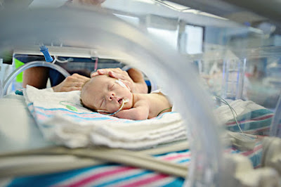 |
| Neonatal intensive care unit |
Diagnostic tests before birth (prenatal care)
Ultrasonography is used before birth to detect many problems and monitor the growth and development of the fetus. Ultrasonography helps doctors determine the sex of the fetus, detect abnormalities in the uterus, detect certain birth defects, and determine the fetus's gestational age. Knowing the fetus's gestational age and birth defects helps doctors anticipate problems that could develop at birth. However, ultrasonography is not 100% accurate. Some babies are born with birth defects that were not detected with ultrasonography.
A triple screen or quad screen is a test done on the mother's blood to measure levels of certain hormones and a protein called alpha-fetoprotein. The test results may be abnormal if the fetus has a genetic abnormality such as Down syndrome (trisomy 21), trisomy 18, spina bifida, or certain other abnormalities. The blood test is done in the first trimester and is often combined with ultrasonography to measure the thickness of the skin folds of the neck of the fetus. Some of the blood tests may be repeated in the second trimester, and this combination of tests is referred to as a sequential screen.
If the results of the screening tests indicate a high risk of abnormalities in the fetus, the mother is offered further testing, most commonly noninvasive prenatal testing (NIPT). NIPT involves genetic testing of the small amounts of fetal DNA that leak from the fetus's circulation into the mother's blood. In some cases, doctors do tests on cells from the fetus using samples taken with a needle from the amniotic fluid (amniocentesis), placenta (chorionic villus sampling), or the umbilical cord (cordocentesis).
Increasingly, NIPT combined with a single blood test of the alpha-fetoprotein level is replacing the triple, quad, and sequential screens. This approach to testing has increased accuracy at an overall lower cost.
Fetal echocardiography, a detailed examination of the heart using a specialized ultrasound device, may be done to detect certain heart defects.
Magnetic resonance imaging (MRI) is now being used to further evaluate some abnormalities in the fetus that are first detected by ultrasonography. MRI may give additional information about an abnormality and can be useful in evaluating treatment options.
Fetoscopy is an invasive test in which doctors insert a small viewing scope (endoscope) into the womb. Early in pregnancy, the scope may be inserted through the mother's cervix. Later in pregnancy, the scope is inserted through a small incision in the mother's abdomen and then another incision in the uterus. The scope lets doctors take a direct look at the placenta and fetus to identify (and sometimes treat) disorders in the fetus.
Diagnosis after birth
After birth, nurses and doctors do a routine physical examination, measure oxygen levels in the blood, and do routine screening tests. Additional tests, including blood tests, X-rays, ultrasonography, and others, maybe done when babies have specific problems or have abnormal findings on the routine tests.
Depending on gestational age, newborns are classified as premature, late preterm, full-term, late-term, or postterm.
Newborns are further classified into three groups by how much they weigh compared to other newborns of the same gestational age. The three groups are,
- Small for gestational age (SGA): Less than the 10th percentile of weight, meaning they are among the 9 lowest-weight babies out of 100 born at a particular gestational age
- Large for gestational age (LGA): Greater than the 90th percentile of weight, meaning they are among the 9 heaviest babies out of 100 born at a particular gestational age
- Appropriate for gestational age (AGA): From the 10th to the 90th percentile of weight, meaning they are among the 82 babies in the middle range for weight
The gestational age and weight classifications help doctors determine the risk of various complications. For example, premature and late preterm newborns are at increased risk of breathing problems because their lungs may not be fully developed. Large-for-gestational-age newborns may have more difficult births and be at increased risk of low blood sugar (glucose).


Comments
Post a Comment